|
|
|
This Project "Studies on Clinical and Forensic Toxicology of Bhopal Aerosol Disaster", constitutes the following distinct areas of research :
Autopsy Findings
Within few hours of the ghastly Gas (now identified as AEROSOL) Tragedy, people started dying. Dr. Varadarajan also agrees toxic chemicals, but never considered change of the title "Gas". From the afternoon of 3rd December, 1984, autopsy work was started by Prof. Heeresh Chandra assisted by the medical/scientific staff of the Medicolegal Institute and the Department of Pathology, Gandhi Medical College, Bhopal. On the 13th December, 1984, ICMR team from the Institute of Pathology, including Dr. S. Sriramachari and Dr. H.M.K Saxena joined Prof Heeresh Chandra. They actively participated not only in the autopsy study but in the subsequent toxicological investigations stemming out of the former.
COMPARATIVE AUTOPSY FINDINGS OF 1985 TO 1987Comparative qualitative and quantitative autopsy findings are as summarised below :
BRAIN
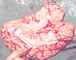
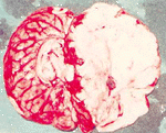
The overall picture in relation to brain weight did not show any marked increase in both the sexes with the subsequent passage of time after 1984. Initial haemorrhagic condition in meaning and later on significantly decreased in the following years.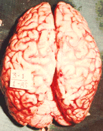
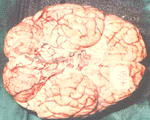
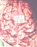
Brain 28 Brain 29 Brain 30
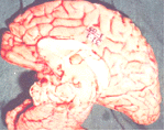

Brain 31 Brain 32 Brain 33
Brain 34
Photograph 28 to 34 Brain surfaces showing stasis in blood vessels, which is charectorised by the absence of venous coloration. The medial surface of the cerebrum shows the same features with generalised oedema. The vessels have become very prominent with increased viscosity resulting in the congestion and pattern appearances. Softening of the white matter was a common feature. These photograph relate to nearly 12 weeks period from the day of disaster.
TRACHEA
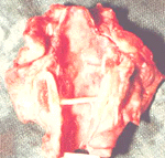
Trachea showing heavy congestion.
LUNGS
As observed in 1984 cases lungs weight were above normal in both the sexes. It was continued till first quarter of 1985 then it became normal. But after the third quarter of 1985 till 1987 in most of the cases it was above normal. Pulmonary oedema, congestion and haemorrhage were the prominent findings of 1984, also observed in less number of cases of 1985 and these findings were constantly seen in 1986 as well as 1987 cases. Pleural thickening was a noted feature in 1985 cases. Bronchitis and pulmonary consolidation were present in higher number of cases of 1985 as compared to 1984 but in the subsequent years these were present in few cases only.
. 

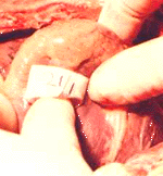
A marked increase in heart weight was observed in both the sexes. During the initial days the cases showed near normal but later on since 11th December 1984 till the end of the month it was above normal and there after it was within the normal range up to June 1985. After July till 1987 a significant increase in weight was observed in most of the cases.
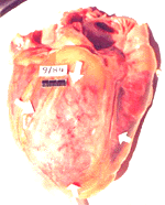
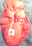
 Heart - 4
Heart 1- first autopsy of 21st December
showing apical petechial haemorrheges
Heart 2- Another heart 6 days after the exposure showing haemorrhages myocardium with petechial haemorrhages and also focal haemorrhages.
Heart 3- External surface of the heart showing generalised haemorrhage with scattered patechial haemorrhages
Heart 4- Haemorrhages seen on the surface of the Heart with coagulated blood in the great blood vessels as cast.
LIVER
Liver constantly showed above normal values of weight in 1984 cases. But since 1985 it was within the normal range in both the sexes. Congestion was prominent feature of 1984 cases which was also continued in most of the cases of 1985, 86 and 87. Liver consistency was soft in most of the cases of the same time.
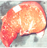
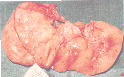
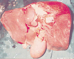
Liver 23 Liver 24 Liver 25
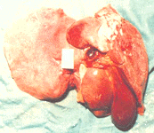
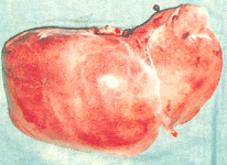
Liver 23 - 27 The external surface of the liver showing petechial and generalised haemorrhages with distended gall bladder - a common finding, and having a ectric tinge in the cut surface.
SPLEEN
In the initial phase of 1984 spleen showed nearly normal values of weight but since 16th December 1984, in males there was a rapid change in weight observed till the end of month and it became normal during mid of 1985. Congestion and softening observed in some cases.
STOMACH

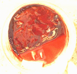

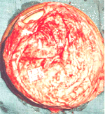
Stomach-2 Hour glass, haemorrhagic appearance of the stomach contents clearly demarcated.
Stomach -3 shows thoracic and abdominal cavity vessels heavily congested with arcades and liver, spleen, showing haemorrhagic surfaces.
Stomach -4 shows the entire thoracic and abdominal cavity with chocolate like liver due to haemorrhage, generalised haemorrgage in the lungs and congestion in the vessels of the intestine
KIDNEYS Kidneys showed constantly much variation in weight, it was more prominent in females and always above normal values with slight increase during December 1984. Congestion and softening continued till 1987 in most of the cases.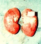
AUTOPSY FINDINGS OF DECEMBER 1984 CASES (SHOWN IN PARENTHESIS)
AUTOPSY FINDINGS OF DECEMBER 1985 CASES (SHOWN IN PARENTHESIS)